
Page 50 - _NIPER-G Annual Report 2019-201
P. 50
Research Area-1
Abietic acid attenuates RANKL induced osteoclastogenesis and inflammation
associated osteolysis by inhibiting the NF‐KB and MAPK signaling
Osteoporosis is a major debilitating cause of fractures and decreases the quality of life in
elderly patients. Bone homeostasis is maintained by bone forming osteoblasts and bone
resorpting osteoclasts. Substantial evidences have shown that targeting osteoclasts using
natural products is a promising strategy for the treatment of osteoporosis. In the current study,
we investigated the osteoprotective effect of Abietic acid (AA) in in vitro and in vivo models of
osteolysis. In vitro experiments demonstrated that, AA suppressed receptor activator of
nuclear factor-kappa B ligand (RANKL)-induced osteoclastogenesis and F-actin ring formation
in a concentration dependent manner. Mechanistically, AA abrogated RANKL-induced
phosphorylation of IKKα/β (ser176/180), IkBα (ser 32), and inhibited the nuclear translocation
of NF-κB.We also found that, AA attenuated the RANKL-induced phosphorylation of MAPKs
and decreased the expression of osteoclast specific genes such as TRAP, DC-STAMP, c-
Fos, and NFATc1. Consistent with in-vitro results, in vivo Lipoploysaccharide (LPS)-induced
osteolysis model showed that AA inhibited the LPS-induced serum surge in cytokines TNF-α
and IL-6. μ-CT analysis showed that AA prevented the LPS-induced osteolysis. Furthermore,
histopathology and TRAP staining results suggested that AA decreased the number of
osteoclasts in LPS-injected mice. Taken together, we demonstrated that the osteoprotective
action of AA is coupled with the inhibition of NF-κB and MAPK signaling and subsequent
inhibition of NFATc1 and c-Fos activities. Hence, AA may be considered as a promising drug
candidate for the treatment of osteoporosis.
Thummuri D, Guntuku L, Challa VS, Ramavat RN, Naidu VG. Abietic acid attenuates RANKL
induced osteoclastogenesis and inflammation associated osteolysis by inhibiting the NF‐KB
and MAPK signaling. Journal of cellular physiology. 2019 Jan;234(1):443-53.
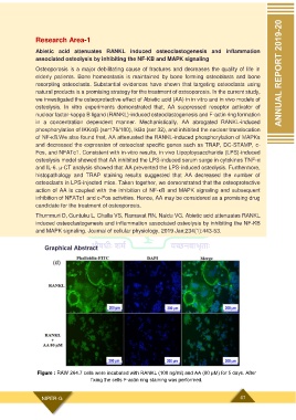
Graphical Abstract
Figure : RAW 264.7 cells were incubated with RANKL (100 ng/ml) and AA (80 μM) for 5 days. After
fixing the cells F-actin ring staining was performed.
NIPER-G 47